



There is an important distinction between showing that an experimental condition can produce an effect and showing that the condition caused the effect in another experiment.
That distinction is at the center of SARS COV 2 ISOLATION PROTOCOL CONTROL EXPERIMENTS, published by Jamie Andrews on September 27, 2025.
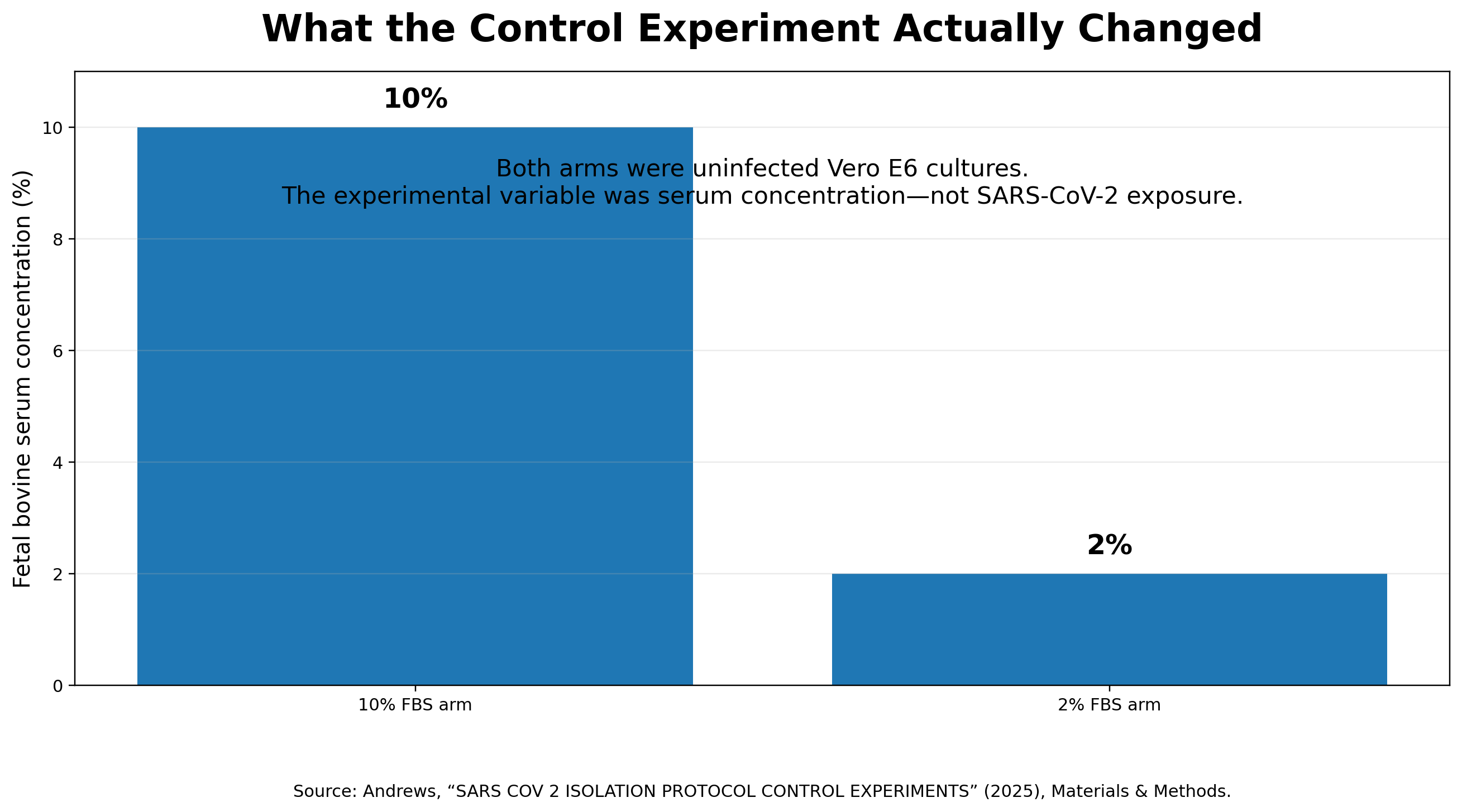
The experiment compares uninfected Vero E6 cells cultured under two serum conditions: 10% fetal bovine serum (FBS) and 2% FBS. The lower-serum cultures grew substantially less and developed morphological changes including rounded cells, granularity, vacuole-like structures, debris and signs of cellular stress.
I am not going to argue that this observation is fake. In fact, doing so would weaken the critique.
Culture conditions can affect Vero-cell growth and survival. In a Vero-cell bioreactor study comparing serum-containing with serum-free medium, the serum-free condition produced a lower maximal viable-cell density and earlier evidence of cell death; apoptosis was detected among detached cells.
That study did not test 10% versus 2% FBS, so it should be treated as background evidence only, not as a replication of Andrews’s experiment.
The legitimate lesson is:
Cell morphology alone can be nonspecific, and culture conditions must be controlled when interpreting cytopathic effects.
That is a useful point. But the Substack article goes much further.
It concludes that serum reduction itself causes the cytopathic effects attributed to viral infection, that cell-culture virus isolation is therefore invalid, and that downstream biochemical assays used to detect viruses are consequently invalid as well.
Those conclusions require evidence that this experiment does not provide. And that is where the argument breaks down.
What the experiment actually shows
The strongest version of the authors’ result is straightforward.
Take otherwise uninfected Vero E6 cultures and alter their serum conditions. Their growth and morphology can change. The contract laboratory’s observations support at least part of that proposition.
The lower-serum cultures showed persistently lower confluence. By later observations, technicians described dense or granular cytoplasm, vacuole-like reflective structures, rounded cells, debris, and cells that might be dying.
That is meaningful.
But there is an important detail that gets lost when the experiment is subsequently summarized as having reproduced viral CPE.
The laboratory’s descriptions are considerably more cautious than the article’s ultimate interpretation.
For several days, even cells in the 2% FBS condition were described as having intact membranes, remaining attached and showing no obvious detachment. On day five, the laboratory characterized the culture as showing some sign of stress, with possible dying cells or debris.
The later classification of these observations into multiple categories of viral CPE is an interpretation by the study authors.
The article itself acknowledges that some claimed morphologies, including syncytia and plaque formation, were not explicitly identified by the laboratory technician. The authors nevertheless interpreted them as being present.
That distinction matters.
The experiment provides evidence of serum-associated differences in cell growth and morphology.
It does not independently establish that every morphology later labeled as CPE was actually present, much less that those morphological changes are equivalent to the complete phenotype seen during a productive SARS-CoV-2 infection.

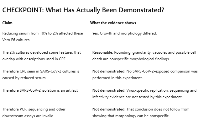
That is the essential distinction. The control experiment establishes a possible confounder. It does not establish that the confounder explains the phenomenon being challenged.
The missing comparison
Suppose changing the nutrient conditions in a cell culture makes some cells round up, grow slowly, become granular, or die.
That immediately raises a sensible question:
Could culture conditions contribute to morphology that might otherwise be interpreted as CPE?
Yes.
But answering the stronger causal question requires comparing an appropriately matched mock condition with a virus-exposed condition while keeping the relevant culture conditions comparable.
Andrews’s experiment does not do that. It contains no SARS-CoV-2-exposed arm. It compares an uninfected 10% FBS culture with an uninfected 2% FBS culture.
That design is useful for asking what changing the serum concentration does to uninfected Vero E6 cells. It cannot determine how much of the effect in an actual SARS-CoV-2 isolation experiment is attributable to serum concentration and how much is associated with the inoculated clinical material and subsequent viral replication.
Calling the experiment a “true negative control” therefore needs qualification.
It is a useful serum-control experiment.
It is not, by itself, a matched mock-versus-virus experiment in which the relevant handling and culture conditions are held constant while viral exposure is the key difference.
That becomes crucial when the argument moves from:
“Reduced serum can produce some overlapping morphology.”
to:
“Reduced serum is the cause of the CPE attributed to the virus.”
The second proposition requires evidence that the first does not supply.
Published virology guidance does not treat CPE alone as definitive
Andrews’s conclusion states that the American Society for Microbiology treats the observation of “any CPE morphology” as sufficient to indicate viral presence.
The ASM protocol is more qualified than that characterization suggests.
ASM explains that some viruses produce characteristic cytopathic effects and says that the microscopic appearance of CPE from some cytocidal viruses may be sufficiently characteristic to allow provisional identification of an unknown virus.
“Provisional” matters. Morphology can provide evidence and help guide identification without being definitive by itself.
FDA guidance for respiratory viral panel submissions is more explicit still. When viral culture is used, FDA recommends virus-specific identification in addition to CPE and states that CPE alone may not provide accurate viral identification.
Banerjee and colleagues made the same distinction in their SARS-CoV-2 isolation study: “The presence of CPE alone does not indicate successful isolation of a coronavirus.”
So Andrews’s control experiment supports a legitimate caution: nonspecific culture conditions can complicate interpretation of cell morphology.
What it does not establish is that conventional viral identification rests on treating any morphological change as conclusive proof of a specific virus.
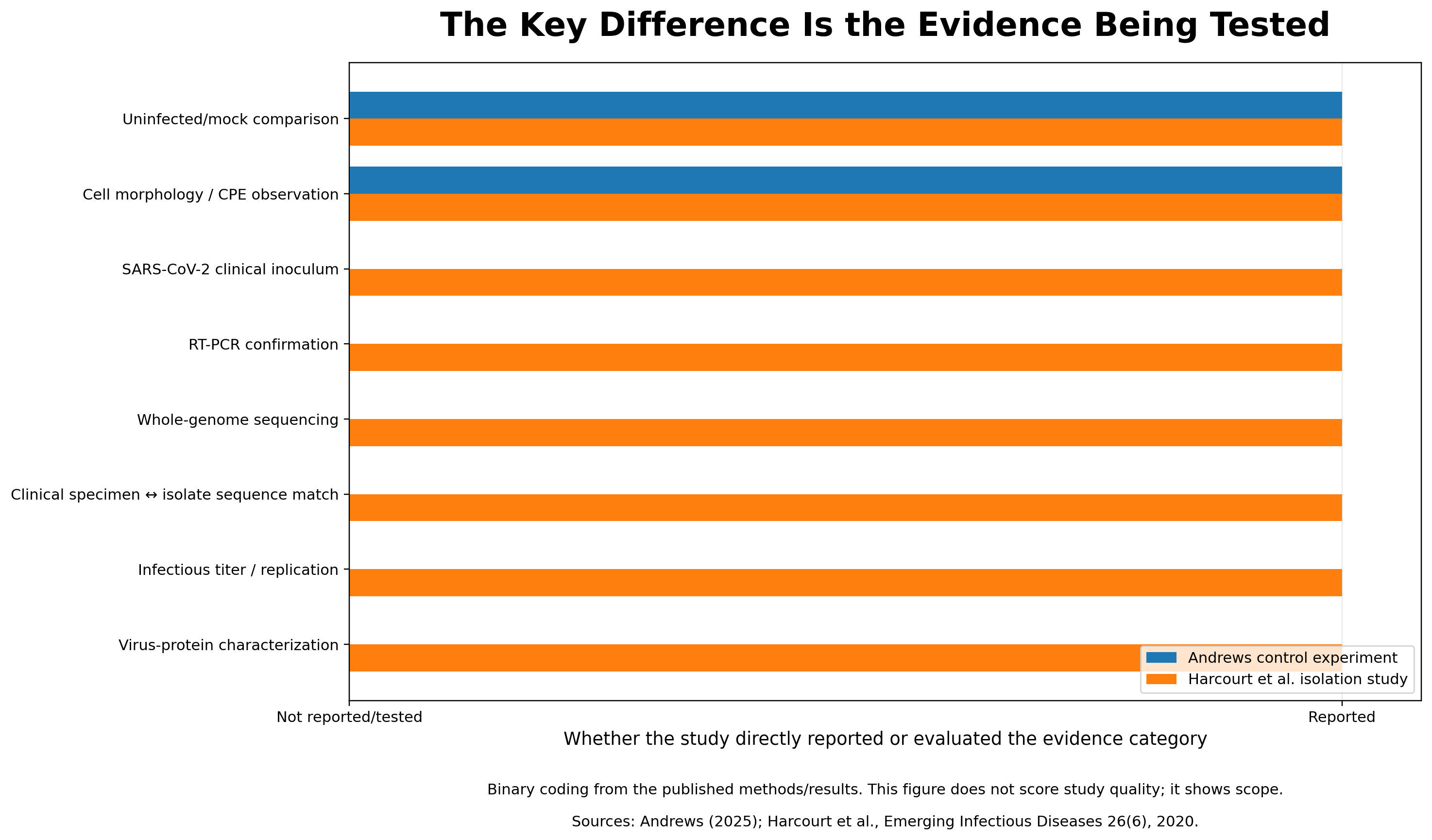
What happens when actual SARS-CoV-2 isolation studies are examined?
This is where the serum-only explanation meets evidence that is not reducible to cell morphology.
Consider the work of Harcourt and colleagues on the first U.S. COVID-19 patient, published in Emerging Infectious Diseases in 2020.
The researchers reported mock-infected cultures alongside cultures inoculated with the patient’s nasopharyngeal and oropharyngeal specimens. CPE was not observed in the mock culture and was observed after inoculation with the clinical specimens.
But the identification did not stop there.
They performed confirmatory RT-PCR on culture lysates, tested the isolates for 33 additional respiratory pathogens, and performed whole-genome sequencing.
Most importantly for the present argument, the genomes obtained from the cultured nasopharyngeal and oropharyngeal isolates were identical to each other and showed 100% identity with the corresponding sequence obtained from the patient’s clinical specimen.
The isolate was also passaged and studied with infectious-titer assays and multistep replication measurements. In Vero cells, infectious titers rose rapidly after inoculation and reached high levels.
Those measurements are not all logically independent of one another, but neither are they simply different names for cell rounding. They include morphology, virus-specific nucleic-acid detection, genome sequence, infectious titer, and replication kinetics.
A serum-associated change in cell shape does not, by itself, account for why a SARS-CoV-2 genomic sequence independently detected in the patient specimen was recovered from the cultured isolate while infectious titers increased after inoculation.
That is the problem for a serum-only explanation: it addresses one possible source of nonspecific morphology, but not the rest of the isolation evidence.
What about contagion?
Another objection sometimes raised is that SARS-CoV-2 has never been shown experimentally to pass from an infected host to a previously uninfected host.
That blanket claim is not supported by the experimental literature.
Dowall and colleagues used the SARS-CoV-2 Victoria/01/2020 strain in a hamster transmission model. Donor hamsters were inoculated with the virus, while naïve recipient hamsters were housed in physically separate adjacent cages connected by directed airflow; the recipients themselves were not experimentally inoculated.
SARS-CoV-2 RNA was subsequently detected in throat swabs from 14 of 16 recipient animals. Clinical signs developed in recipient animals, and histopathological examination found pneumonia in 10 of 16 recipients.
There is an important limitation worth stating explicitly: the Dowall study did not report recovery of live virus from recipient throat swabs. Its evidence in the recipients consisted of viral RNA, clinical findings, and histopathology. It should therefore be cited for transmission-associated infection and disease in this model, not as evidence that infectious virus was re-isolated from those particular recipient swabs.
Other animal studies provide complementary evidence. Sia and colleagues reported efficient SARS-CoV-2 transmission from inoculated to naïve hamsters by direct contact and by aerosols; naturally infected animals developed weight loss and neutralizing antibodies.
In ferrets, Richard and colleagues demonstrated transmission by direct contact and through the air, and reported that infectious virus was isolated from all animals that tested positive.
The 2022 Nature Medicine human challenge study addresses a related but different question. It was not a person-to-person transmission experiment. Thirty-six healthy adults aged 18–29 with no evidence of previous infection or vaccination were deliberately inoculated with a defined SARS-CoV-2 preparation; 34 were included in the per-protocol analysis and 18 became infected.
Viral load rose rapidly, viable virus was recoverable from the nose, and most infected participants developed mild-to-moderate symptoms. The study found no clinical or radiological evidence of pulmonary disease in these carefully selected young adults.
So the evidence should be described precisely:
Controlled human infection has been demonstrated, and experimental host-to-host transmission has been demonstrated independently in multiple animal models.
Those are different experimental questions. Neither should be overstated, but neither can fairly be summarized as “there has never been a successful contagion study.”
What about demanding a live video of the virus entering a cell and replicating?
That is not a scientific criterion for establishing the existence or replication of a virus.
Different techniques answer different questions. Conventional transmission electron microscopy provides high-resolution structural information but generally examines fixed specimens, so it is not designed to provide a continuous live movie of an entire infection cycle.
Live-cell systems can visualize infection and spread over time. In a 2024 Nature Communications study, researchers used a fluorescent reporter virus derived from SARS-CoV-2 USA/WA1-2020 to image infected differentiated human bronchial epithelium longitudinally for up to 12 days. The fluorescent signal marked infected cells and allowed infection foci and their movement through the tissue to be followed over time.
Earlier work in the Journal of Virology used a reporter-cell system during SARS-CoV-2 infection and performed live imaging every 10 minutes for 18 hours, observing a time-dependent reporter response in infected cells.
Direct structural observation is also available, although it is not live imaging. Klein and colleagues used in situ cryo-electron tomography of SARS-CoV-2-infected cells to visualize replication-associated double-membrane vesicles, RNA filaments, virion budding sites, spike trimers, viral ribonucleoprotein complexes, intracellular virions, and extracellular virions.
Those studies should not be oversold. They are not a literal camera view of one unmodified virion being followed continuously through attachment, entry, uncoating, genome copying, assembly, and release.
That is not their purpose.
Replication is instead evaluated through converging measurements appropriate to different stages of the process—for example, recovery of infectious material, changes in infectious titer over time, virus-specific nucleic-acid detection, genome sequencing, viral protein expression, replication kinetics, serial passage, and infection of additional susceptible cells.
A demand for one cinematic image sequence does not negate those measurements. The relevant question is whether the individual methods are properly controlled and whether their results converge.
Not every SARS-CoV-2 isolation protocol used the same serum regimen.
Andrews’s article makes a broad claim that documented SARS-CoV-2 isolations reduced infection medium to 2% or even 1% FBS.
The Harcourt protocol does not fit that description neatly.
Harcourt and colleagues report maintaining Vero cultures in medium containing 5% or 10% FBS. During the initial isolation procedure, the Vero cells were resuspended in DMEM containing 10% FBS before being added to the clinical-specimen dilutions.
There is an important precision point here: because that cell suspension was mixed with specimen and serum-free diluent already present in the wells, the final FBS concentration in every well should not be described simply as 10%.
But the published method also does not describe the specific switch to a 2% FBS infection medium tested in Andrews’s control experiment.
That is enough to show that the serum-regimen claim is not universal. SARS-CoV-2 isolation protocols have not all used the same serum regimen, and the effect of a particular 10%-to-2% switch cannot simply be generalized to every isolation protocol.
What about studies that actually did use 2% FBS?
Banerjee and colleagues are particularly relevant because their 2020 SARS-CoV-2 isolation method explicitly states that, after the initial inoculation period, the inoculum was replaced with DMEM containing 2% FBS and TPCK-treated trypsin.
The paper also shows mock-inoculated Vero E6 cultures alongside cultures inoculated with clinical specimens, and the specimen-inoculated cultures developed extensive CPE relative to the mock cultures.
There is, however, a limitation that should not be glossed over: the methods do not separately spell out every reagent and concentration used in the mock arm. For that reason, this paper should not be presented as definitive proof that the mock cultures were matched to the infected cultures in every medium component, including the exact 2% FBS condition.
Fortunately, the argument does not need that claim.
Banerjee and colleagues themselves emphasized that CPE alone was insufficient for identification. They transferred supernatant from the clinical-specimen cultures to fresh Vero E6 cells, confirmed SARS-CoV-2 RNA in culture supernatant by RT-PCR, generated approximately 94%-complete SARS-CoV-2 genome sequences at very high sequencing depth, measured infectious titers, and went on to characterize replication competence.
The paper also reports sequence substitutions shared with direct sequencing of clinical isolates, but that particular comparison refers in part to unpublished data. I therefore would not use that comparison as a central piece of the argument.
The stronger published point is simpler: even in an isolation protocol that used 2% FBS, the investigators did not treat cell morphology as sufficient evidence of viral identity. They combined culture observations with molecular identification, sequencing, infectivity, and replication measurements.
This is where the causal argument fails
The control-study argument effectively proceeds this way:
Reduced serum can produce cellular abnormalities.
Some of those abnormalities resemble features that can occur during viral CPE.
Some virus-isolation protocols use reduced-serum media.
Therefore, reduced serum causes the CPE attributed to viruses.
Therefore, the claimed virus was not actually isolated.
Therefore, molecular tests built around that virus are invalid.
The first three points support a hypothesis worth testing.
They do not establish the remaining conclusions.
To show that serum reduction causes the findings attributed to SARS-CoV-2, the evidence would have to distinguish the effects of the culture condition from the additional effects associated with the clinical inoculum and with recoverable, replicating infectious material.
Andrews’s experiment never performs that comparison.
That is the missing causal link.
The electron-microscopy criticism is partly right
The article also argues that uninfected cells can contain structures resembling particles that have sometimes been identified as coronaviruses under transmission electron microscopy.
That concern is legitimate.
Electron-microscopy specialists Cynthia Goldsmith, Sara Miller, and colleagues cautioned during the pandemic that normal cellular structures—including coated vesicles and multivesicular bodies—can be mistaken for coronavirus particles.
The supported conclusion is therefore narrow but important:
Ambiguous TEM morphology should not be used by itself to identify SARS-CoV-2.
But resemblance is not identity.
Finding a vesicle-like structure in an uninfected culture that resembles a published viral image does not establish that the published structure was also a cellular vesicle, nor does it invalidate virus identification supported by additional molecular, genomic, and infectivity evidence.
This is also why the in situ cryo-electron tomography work matters. In infected cells, investigators have visualized not merely isolated round particles but replication-associated compartments, viral RNA-containing structures, budding events, spike trimers, viral ribonucleoprotein complexes, and assembled virions in their cellular context.
Morphology is strongest when it is interpreted together with the rest of the evidence rather than treated as a stand-alone verdict.
“Isolation” does not mean “chemical purification”
Another major point of disagreement concerns the word isolation.
Andrews argues that unless researchers first obtain a purified, distinct biological particle, subsequent identification lacks a valid benchmark.
That is not the conventional meaning of virus isolation in clinical virology.
CLSI’s own terminology database defines viral culture as the infection of cell cultures by viable infectious viral particles, usually resulting in viral replication, and lists “viral isolation” as an alternate term.
A major review in Clinical Microbiology Reviews likewise repeatedly uses “virus isolation in cell culture” to describe recovery of viable virus in susceptible cells followed by identification and confirmation.
This terminology does not mean purification is useless.
Purification can be essential for particular structural, biochemical, or analytical questions. It is simply a different operation from recovering and propagating a viable virus isolate in susceptible cells.
The distinction matters because otherwise the argument becomes circular: redefine “isolation” to mean “chemically purified virions only,” then declare every conventional cell-culture isolation invalid because it does not satisfy that redefinition.
A fair critique should instead evaluate conventional virus isolation by the criteria the method actually claims to satisfy: recovery of infectious material, reproducible propagation in susceptible systems, appropriate controls, and identification by suitable independent methods.
A note about the CLSI claim
The control article invokes CLSI M41 as support for the proposition that infection media are supposed to be reduced to roughly 1–3% FBS.
This point needs careful wording.
The public CLSI page and publicly available sample confirm that M41 is a 2006 viral-culture guideline covering cell-culture selection and maintenance, quality control, culture-medium preparation, specimen handling, isolate identification, and interpretation. CLSI currently lists it as an archived document that remains technically valid.
The publicly accessible material I could verify does not expose enough of the full standard to establish whether the cited 1–3% FBS recommendation appears in the precise form Andrews describes.
So I would not claim that M41 contains no such recommendation.
But the public material also does not support presenting a specific low-serum concentration as a universal rule governing every virus-isolation protocol.
And even if the full M41 document recommends reduced-serum maintenance medium for particular applications, that would establish a culture practice—not the causal proposition that reduced serum is therefore responsible for the virus-specific findings reported in SARS-CoV-2 isolation studies.
The relevant experimental question remains:
What happens to appropriately matched inoculated and mock-inoculated cultures under comparable conditions, and what independent evidence identifies the recovered agent?
Modern SARS-CoV-2 characterization is explicitly multi-method
This point has become even clearer with time.
ECDC’s updated 2026 handbook for SARS-CoV-2 characterization does not describe identification as “look for CPE and stop.”
It covers cell culture alongside plaque assays, viral titration, antibody-based detection, sequencing and PCR, and specifically emphasizes that protocols vary among institutes and sometimes require adaptation.
That is important because the control article treats one culture condition as though it were the single foundation beneath SARS-CoV-2 identification.
It isn’t.
The evidentiary picture is distributed across independent techniques.
That is exactly what one should want.
The largest unsupported leap: “therefore downstream assays are invalid”
The article’s final step is its broadest.
It argues that if cell-culture isolation is invalid, downstream molecular and biochemical assays must also be invalid because no properly isolated benchmark exists.
But Andrews’s experiment does not test those assays.
It does not test sequencing specificity.
It does not test PCR specificity.
It does not show SARS-CoV-2 sequences arising in its uninfected cultures.
It does not demonstrate recoverable infectious virus in an uninoculated culture.
It does not show the patient-associated viral genome appearing reproducibly as a consequence of reduced serum.
Nor does the experiment address the Harcourt result in which the sequence recovered from the cultured isolate matched the corresponding sequence obtained directly from the clinical specimen.
That observation matters because the sequence was documented in the patient specimen independently of the subsequent culture result. The cultured isolate then yielded the corresponding sequence while infectious-virus measurements showed replication in susceptible cells.
One can raise separate questions about PCR design, sequencing, contamination control, or assay validation. Those questions should be evaluated on their own evidence.
A serum-only experiment that measures cell morphology does not, by itself, invalidate them.
What would make this control experiment much more consequential?
This is the scientific question that matters.
Imagine that an appropriately matched mock control, subjected to the same relevant culture and handling conditions, repeatedly and reproducibly produced not merely similar-looking cellular damage but also the same SARS-CoV-2-specific molecular signal, a corresponding viral genome, and serially transferable or quantifiable infectious activity attributed to the supposed isolate.
That would be a profoundly important result.
It would directly challenge the specificity of the isolation process and demand an explanation.
But that is not what this experiment reports.
It reports morphological changes in uninfected Vero E6 cells exposed to different serum conditions.
Interesting?
Yes.
Worth considering when interpreting CPE?
Absolutely.
Evidence that morphology should not stand alone?
Certainly.
Evidence, by itself, that SARS-CoV-2 isolation has been disproven?
No.
The strongest conclusion is also the narrowest one
There is no need to dismiss Andrews’s experiment.
Its strongest observation can simply be accepted:
Reducing serum from 10% to 2% altered the growth and morphology of these uninfected Vero E6 cultures, and several observed features overlap with morphological categories that can also occur during viral CPE.
That supports careful controls.
It supports caution about identifying infection from morphology alone.
And it supports skepticism toward any experiment that treats a nonspecific visual change as sufficient proof of viral identity.
But that is not where the evidence in SARS-CoV-2 isolation studies ends.
Andrews’s experiment never exposes cells to SARS-CoV-2. It therefore does not measure the difference between serum-associated cell stress and the additional effects associated with a SARS-CoV-2-containing clinical inoculum.
Its design also does not test or account for:
recovery of replication-competent infectious material from clinical specimens;
rising or quantifiable infectious titers;
virus-specific molecular detection;
genome sequencing;
correspondence between sequences recovered directly from clinical specimens and cultured isolates;
serial passage into additional susceptible cells; or
experimental infection and host-to-host transmission evidence from other study designs.
That is the central problem.
The experiment identifies a plausible confounder for interpreting cell morphology. It then treats the existence of that confounder as proof that the confounder explains the entire isolation result.
Those are not the same thing.
The strongest criticism of the study is therefore not that its cell-stress observation must be false. It is that the conclusion extends far beyond what the experiment was designed to test.
When the Conclusion Is Built Into the Standard
Some critics begin by declaring that only one particular method can count as valid evidence. Once that rule is adopted, every other line of evidence is rejected before it is examined. That is the central flaw: the conclusion is built into the standard being imposed.
Disagreeing with the accepted evidentiary framework does not demonstrate that the evidence within that framework is false; it merely shows that the critic refuses to recognize the kinds of converging evidence the field uses. A methodological preference is not, by itself, an experimental refutation.
Resources and Primary Sources
Jamie Andrews — “SARS COV 2 ISOLATION PROTOCOL CONTROL EXPERIMENTS”, September 27, 2025.
The experiment evaluated in this article. It compares uninfected Vero E6 cultures maintained under 10% and 2% FBS conditions and argues that serum reduction can produce morphology interpreted as CPE and invalidate virus isolation.
Harcourt J, Tamin A, Lu X, et al. “Severe Acute Respiratory Syndrome Coronavirus 2 from Patient with Coronavirus Disease, United States.” Emerging Infectious Diseases. 2020;26(6):1266–1273. doi:10.3201/eid2606.200516.
Reports mock cultures, confirmatory RT-PCR, testing for 33 additional respiratory pathogens, whole-genome sequencing, a 100% sequence match between cultured isolates and the corresponding clinical specimen, infectious-virus quantification, and replication studies.
Banerjee A, Nasir JA, Budylowski P, et al. “Isolation, Sequence, Infectivity, and Replication Kinetics of Severe Acute Respiratory Syndrome Coronavirus 2.” Emerging Infectious Diseases. 2020;26(9):2054–2063. doi:10.3201/eid2609.201495.
The isolation method used 2% FBS after inoculation. The paper included mock-inoculated cultures and explicitly stated that CPE alone does not establish successful coronavirus isolation. The methods do not separately itemize every reagent concentration for the mock arm, so this article does not rely on Banerjee as proof of a perfectly matched 2% FBS mock.
American Society for Microbiology — “Cytopathic Effects of Viruses Protocols.”
Describes characteristic CPE as potentially permitting provisional identification of an unknown virus rather than definitive identification from morphology alone.
U.S. Food and Drug Administration — “Respiratory Viral Panel Multiplex Nucleic Acid Assay: Class II Special Controls Guidance.”
States that CPE alone may not provide accurate viral identification and recommends virus-specific identification in addition to CPE when viral culture is used in this regulatory context.
Clinical and Laboratory Standards Institute — “M41: Viral Culture.”
A 2006 guideline covering viral culture, culture-medium preparation and quality control, specimen processing, isolate identification, and interpretation. CLSI lists it as archived but technically valid.
Leland DS, Ginocchio CC. “Role of Cell Culture for Virus Detection in the Age of Technology.” Clinical Microbiology Reviews. 2007;20(1):49–78. doi:10.1128/CMR.00002-06.
A major review describing the conventional use of cell culture for virus isolation and the combination of culture with antigenic and molecular identification methods.
Goldsmith CS, Miller SE, Martines RB, Bullock HA, Zaki SR. “Electron microscopy of SARS-CoV-2: a challenging task.” The Lancet. 2020;395(10238). doi:10.1016/S0140-6736(20)31188-0.
Discusses the difficulty of correctly distinguishing coronavirus particles from normal cellular structures by electron microscopy.
Miller SE, Goldsmith CS. “Caution in Identifying Coronaviruses by Electron Microscopy.” Journal of the American Society of Nephrology. 2020;31(9):2223–2224. doi:10.1681/ASN.2020050755.
Documents the risk of mistaking ordinary cellular structures, including coated vesicles and multivesicular bodies, for coronavirus particles.
Quesney S, Marvel J, Marc A, Gerdil C, Meignier B. “Characterization of Vero cell growth and death in bioreactor with serum-containing and serum-free media.” Cytotechnology. 2001;35(2):115–125. doi:10.1023/A:1017589526145.
Background evidence that serum conditions can affect Vero-cell growth and cell death. This study compared serum-containing with serum-free conditions, not 10% versus 2% FBS.
European Centre for Disease Prevention and Control — “Update of standard laboratory protocols for SARS-CoV-2 characterisation – 2026.”
Describes multiple methods used in SARS-CoV-2 characterization, including cell culture, plaque assays, viral titration, antibody-based methods, neutralization, PCR, and sequencing, and notes protocol variability between institutes.
Killingley B, Mann AJ, Kalinova M, et al. “Safety, tolerability and viral kinetics during SARS-CoV-2 human challenge in young adults.” Nature Medicine. 2022;28:1031–1041.
A controlled human infection study: 36 young adults were inoculated, 34 were included in the per-protocol analysis, and 18 became infected. Viable virus was recoverable from infected participants; the study did not test person-to-person transmission and found no pulmonary disease in these carefully selected volunteers.
Sia SF, Yan L-M, Chin AWH, et al. “Pathogenesis and transmission of SARS-CoV-2 in golden hamsters.” Nature. 2020;583:834–838.
Demonstrated efficient transmission from inoculated to naïve hamsters by direct contact and aerosols. Naturally infected animals developed weight loss and neutralizing antibodies.
Richard M, Kok A, de Meulder D, et al. “SARS-CoV-2 is transmitted via contact and via the air between ferrets.” Nature Communications. 2020;11:3496.
Demonstrated direct-contact and airborne transmission between ferrets and reported infectious virus isolation from all positive animals.
Dowall S, Salguero FJ, Wiblin N, et al. “Development of a Hamster Natural Transmission Model of SARS-CoV-2 Infection.” Viruses. 2021;13(11):2251. doi:10.3390/v13112251.
Used SARS-CoV-2 Victoria/01/2020 in a physically separated airflow transmission system. Viral RNA was detected in 14 of 16 recipients and pneumonia in 10 of 16. The study did not report live-virus recovery from recipient throat swabs.
Becker ME, Martin-Sancho L, Simons LM, et al. “Live imaging of airway epithelium reveals that mucociliary clearance modulates SARS-CoV-2 spread.” Nature Communications. 2024;15:9480. doi:10.1038/s41467-024-53791-4.
Used a fluorescent reporter virus derived from SARS-CoV-2 USA/WA1-2020 and longitudinal live imaging of differentiated primary human bronchial epithelium for up to 12 days, allowing infection foci and viral spread to be followed over time.
Klein S, Cortese M, Winter SL, et al. “SARS-CoV-2 structure and replication characterized by in situ cryo-electron tomography.” Nature Communications. 2020;11:5885. doi:10.1038/s41467-020-19619-7.
Used in situ cryo-electron tomography to directly visualize SARS-CoV-2 replication-associated structures, viral ribonucleoprotein complexes, budding sites, and assembled virions in infected cells. These are cryopreserved structural observations, not live-cell movies.
Pahmeier F, Neufeldt CJ, Cerikan B, et al. “A Versatile Reporter System To Monitor Virus-Infected Cells and Its Application to Dengue Virus and SARS-CoV-2.” Journal of Virology. 2021;95. doi:10.1128/JVI.01715-20.
Used a reporter-cell system for live-cell monitoring of SARS-CoV-2 infection, including imaging at 10-minute intervals over an 18-hour period.